Careful Interpretation Even for Minor Conditions
1:1 Specialist Consultation
Increasing Rate of Breast Conditions and Breast Cancer
Breast cancer is the most common women’s cancer in Korea, and its prevention is growing more important.
Our specialists at MEDIWOMAN Clinic provides specialized care for women’s breast health.
Symptoms of Breast Cancer
                                About 70% of breast cancer symptoms are tumor (bump, lump, nodule),
                                and breast ultrasound and biopsy are performed to determine whether it is malignant (breast cancer) or benign.
However, since symptoms of benign tumors and breast cancer are similar, it is crucial to detect and treat at an early stage.

-
CASE 1

Breast lump(bump)
-
CASE 2

Breast inflammation
-
CASE 3

Breast pain
-
CASE 4

Breast change
-
CASE 5

Skin eczema on breast area
-
CASE 6

Retracted nipple
-
CASE 7

Nipple discharge
-
CASE 8

Armpit lump (bump)
-
CASE 9

Armpit Boil
-
CASE 10

No pain (asymptomatic)
Breast Examination
MEDIWOMAN provides a step-by-step examination to diagnose and treat benign and malignant breast conditions.
                            Dense breasts with abundant glandular tissue are common in Korean women, and mammography, ultrasound,
                            and biopsy should be performed by experienced medical staff for accurate diagnosis.
-
1
Consultation with Director

Breast surgery specialists with over 30 years of clinical experience consult and physically examine to identify main symptoms, medical history, and family history, and provide 1:1 personalized treatment based on ultrasound and mammography results.
-
2
Mammography
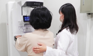
For X-ray imaging, the breast is compressed using mammography device designed for detecting breast microcalcification.
-
3
Breast Ultrasound
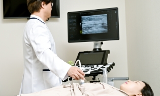
High-resolution ultrasound device is used to diagnose malignant tumor (breast cancer) and benign tumor (bump, lump, nodule) in breasts with complex glandular tissue. Since the device examines deeper tissues, it can identify lesions that may not be detected in mammography.
-
4
Breast Biopsy (Mammotome)
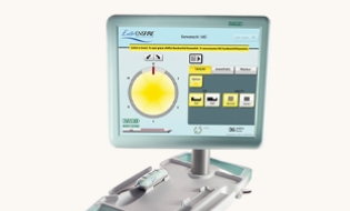
Breast biopsy is a highly accurate test, and it takes a portion of suspected tumor to determine whether it is malignant or benign. For patients with family history of breast cancer, it is recommended to undergo both breast ultrasound and biopsy.
Get Mammography and Breast Ultrasound Together
For More Accurate Breast Cancer Screening
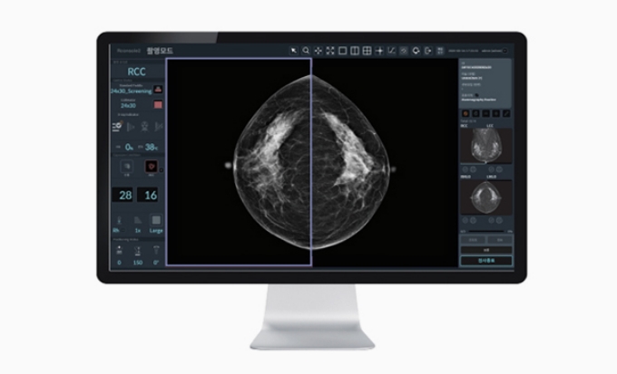
                            Mammography is mainly performed to diagnose breast cancer.
                            However, as many Korean women have dense breasts, it is often difficult to distinguish glandular tissue and fat tissue.
Therefore, it is recommended to get both mammography and breast ultrasound to increase the accuracy.
High-Resolution Breast Ultrasound
Examines deeper tissues with higher resolutions to double-check and identify lesions that are difficult to detect in mammography.
AI-Based Mammography Device
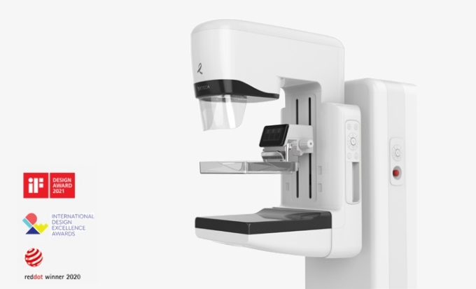
Creates high-resolution images to detect even subtle lesions and uses AI technology to increase the accuracy of breast cancer diagnosis.
Mammotome
Screens Breast Cancer and Removes Lesion Simultaneously
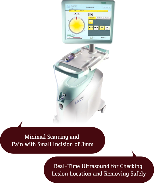
                            Mammotome is also known as a vacuum-assisted breast tumor excision procedure.
                            It checks breast ultrasound in real-time to take sample tissues of breast nodules (lump, tumor) and remove the lesion.
                            Mammotome involves a minimal incision of 3mm, and it minimizes burden of scarring and pain.
                            It also shows high accuracy rate by extracting larger tissue samples compared to a core needle biopsy.
Still, it requires high level of technical expertise, as precise lesion location needs to be checked through ultrasound during procedure.

Mammotome Process
-
1
Highly Accurate
Real-Time Ultrasound
Real-Time Ultrasound Monitoring to Confirm Lesion Size and Location During Procedure.
-
2
Minimum Incision for
Lower Burden of Scarring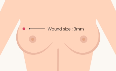
Mammotome is performed with a minimum 3mm-incision to sample breast nodules (lump, tumor) and remove the lesion, and it is followed by careful suture.
-
3
Post-Procedure Care

Patients are discharged on the same day, and get regular follow-up care, such as lesion removal check.
Strengths of Mammotome
Pain
Advanced Technical Skills for
Removing Large or Multiple Tumors
As increasing cases of multiple breast tumors are reported, breast cancer rate is also increasing accordingly.
Mammotome procedure must evolve in response to the changing breast cancer diagnosis.
If necessary, Mammotome procedure should be actively considered for early diagnosis and treatment.
Large Tumors Over 3cm
                                        Real-time ultrasound identifies the exact location of the tumors to
                                        remove them regardless of size.

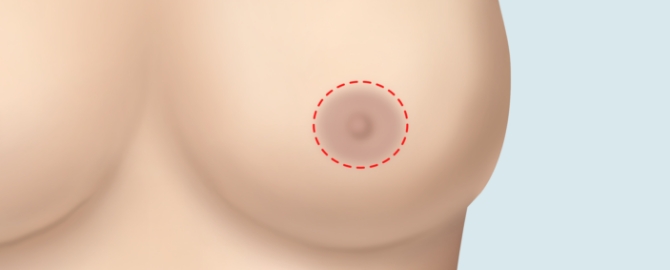
Areolar Approach
                                        An incision is placed along the border of the skin and areola to
                                        make the scarring unnoticeable.
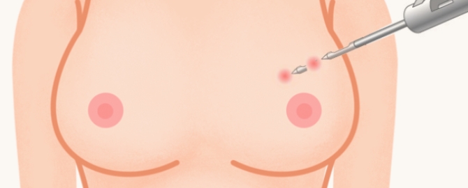
Multiple Tumors
                                        In cases of multiple breast tumors, a procedure is performed through
                                        a single incision area to minimize scarring and pain.
Patients with Breast Implants
                                        For patients with breast implants, Mammotome needle extracts or removes
                                        only tumor tissues, even if located in challenging anatomical areas,
                                        without damaging the implants.